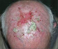
EROSIVE PUSTULAR DERMATOSIS OF THE SCALP
Luciano Schiazza M.D.
Dermatologist
c/o InMedica - Centro Medico Polispecialistico
Largo XII Ottobre 62
cell 335.655.97.70 - office 010 5701818
www.lucianoschiazza.it
Erosive pustular dermatosis of the scalp is a chronic inflammatory disease that usually occurs in elderly individuals in areas with sparse hair.

It was described in 1979 by Pye, Peachey and Burton.
It is characterized by sterile pustules (rarely visualized), erosion, yellow-brown crusts on an atrophic skin of the scalp. The EPD can result in scarring alopecia if not treated.
Clinical findings could suggest the presence of an infection, but signs of infection as swelling, warmth, regional lymphadenopathy are lacking.
Although etiology remains poorly understood, several factors may be related to the pathogenesis of the disease:
-
Atrophied skin, most commonly secondary to actinic damage, (a predisponing condition),
-
Trauma with tissue damage,(precipitating factor). Other insulting causes include herpes zoster, cryotherapy, topical chemotherapy, excisional and grafting surgery, photodynamic therapy, carbon dioxide laser therapy.
Chronic inflammation noted in histology studies suggests that it plays a role in the persistence of the disease because
is always present, moderate to marked,
the disease responds well to anti-inflammatory therapy to topical steroids.
The clinical differential diagnoses include:
-
actinic keratoses,
-
Squamous Cell Carcinoma
-
Pyoderma gangrenosum
-
Pemphigujs fogliaceo,
-
Factitial damage
-
Chronic bacterial or fungal infection.
Other possibilities:
-
Pustular psoriasis
-
Cicatricial pemphigoid,
-
Pemphigus vulgaris,
-
Darier’s disease,
-
Chronic mucocutaneous candidiasis,
-
Dissecting cellulitis,
-
Neutrophilic scarring alopecia
-
Folliculitis decalvans
EPD is a clinical diagnosis of exclusion. A skin biopsy should be perform to confirm, for the diagnosis of the disease, the non specific histopathological findings of the specimen: epidermal atrophy, parakeratotic and ortokeratotic scales, atrophy, inflammatory dermal infiltrate (lymphocytes, neutrophils, plasma cells).
When neutrophils are present subcorneal and spongiform pustules may be noted. Special staining and culture for bacteria and fungi are negative as direct immunofluorescence on biopsy tissue.
The disease has a good response to high-potency topical steroids. Also tacrolimus ointment has been reported to be helpful.
